


LIFTING DOCTOR NOTE
LIMO PLASTIC SURGERY
manager
123times Date 26-07-16 11:42
- Operator : Dr Kwon Young Hun
- Previous surgery history : Facial contouring surgery (3 years ago), liposuction (double chin, cheeks, 1 year ago)
- Significant : A Lt. congenital melanocytic nevus (CMN) is present, spanning approximately 4 cm of skin including the earlobe and jawline area.
- Age : 20s Gender : Female Japanese Patient
Sedation (sleep anesthesia) was administered, and the incision sites were additionally designed.
After local anesthesia, slit incisions were made, and 20cc of tumescent solution was injected (both sides).
Following incision at the designated sites, thin skin flap elevation was performed while preserving the SMAS. Severe scarring was observed around the zygoma area, mandible margin, and neck, presumed to be the result of the previous face contouring surgery and liposuction (cheeks, double chin); dissection was carried out with maximal effort to avoid SMAS injury. (Aside from the SMAS injury near the anterior ear caused by the previous zygoma reduction, the SMAS was intact.)
After completing the dissection, bleeding control was performed, followed by SMAS dissection.
Given the scarring around the zygoma from the previous contouring surgery, the Extended High SMAS technique was used.
Due to adhesions from the previous zygoma (cheekbone) surgery, severe adhesion was present around the zygomatic major area. The zygomatic ligament was identified and released; the maxillary ligament and mandibular ligament (retaining ligaments) were also identified and dissected/removed. Adhesion in the lower area was less severe than at the zygoma.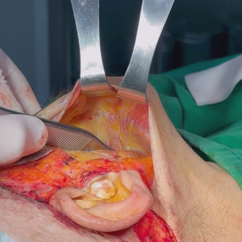
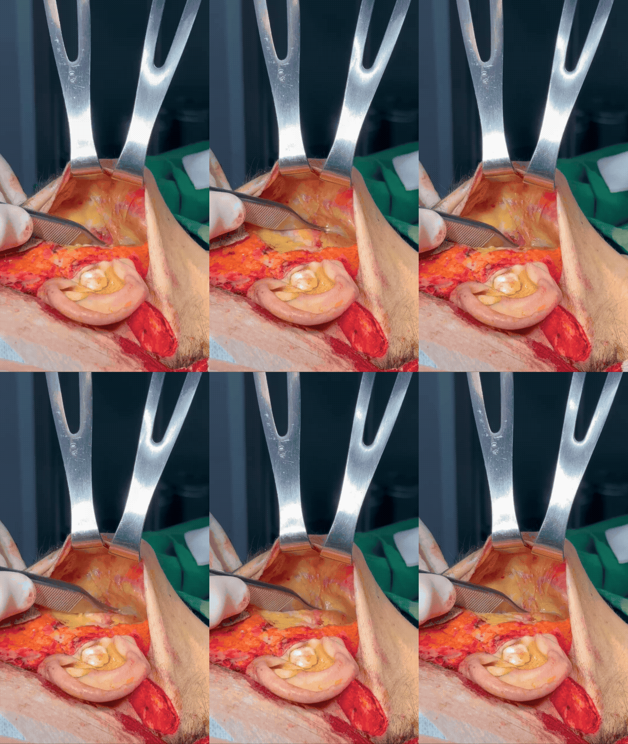
▲ (Image showing the anatomy before the retaining ligaments were released)
After tension-free SMAS elevation, SMAS fixation was performed; the same procedure was carried out on the contralateral side. Since adhesion was less severe on the left side, to remove the Lt. CMN as much as possible, the SMAS was dissected in a tension-free manner, and the excess SMAS was trimmed, and fixation was performed.
Before skin closure, liposuction of the lateral cheek and double-chin line was performed; the fat compartment did not contain a large volume due to the previous liposuction, and the procedure was carried out to maximize re-adhesion (fixation) in that area.
After bleeding control on both sides, the excess skin was removed. For the Lt. CMN, approximately 90% was removed (excluding the earlobe), leaving about 2–3 mm at the incision site. (As the patient is young, skin closure was also performed tension-free with consideration for the final scar outcome.)
After insertion of the Hemovac drain, double-layer skin closure was performed. No specific abnormalities occurred during the surgery.